Learning about Pelvic Ultrasound: An Important Diagnostic Modality
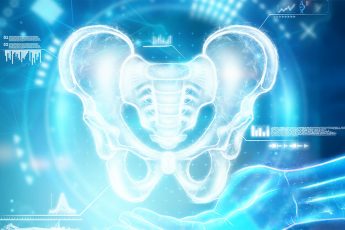
Medical imaging is extremely important when it comes to assessing musculoskeletal disease, enabling practitioners to explore structures inside the body without ever actually being inside. One of the most common imaging tools is the pelvic ultrasound, an innocent and harmless procedure that uses high-frequency sound waves to visualize the bones and organs of the pelvic area.
Although ultrasounds in the pelvic area are usually linked to reproductive issues, they can also be of great use in identifying orthopedic issues in the bones, joints, and the soft tissues in the pelvis. Here at Adam Vital Hospital, we have state-of-the-art ultrasound equipment that assists our orthopedics and sports medicine divisions in assessing hip and pelvic-related conditions.
What is a Pelvic Ultrasound?
A pelvic ultrasound employs sound waves to move within the body and create real-time images of pelvis structures, soft tissues, muscles, ligaments, and bones. Unlike CT scans or X-rays, ultrasound does not employ radiation and hence is less intimidating as a repeated test, and also for subjects of any age group.
For musculoskeletal conditions, pelvic ultrasound images joint structures, bony shapes, and adjacent soft tissues in patients with pain in the pelvis, hips, or restrictions in mobility.
Musculoskeletal Pathology That May Require a Pelvic Ultrasound
Hip Dysplasia
Hip dysplasia is a condition where the hip socket does not fully cover the top of the upper thighbone. Hip dysplasia can cause dislocations, abnormally worn parts, and early arthritis if left undetected and untreated. Ultrasound of the pelvis is the main imaging method used in infants and toddlers to detect developmental dysplasia of the hip (DDH).
In adults, ultrasound is helpful in the evaluation of adjacent muscles and ligaments, particularly for postoperative evaluation or injury.
Injuries to the Tendon and Muscle
Ultrasound of the pelvis facilitates imaging of the muscle tears, tendon injuries, and soft tissue inflammation of the pelvic girdle. Competitive athletes and physically active individuals may be evaluated using ultrasound when they present with complaints of groin pain or pelvic strain.
This imaging technique enables the doctor to see minute injury that is not visible on X-rays, particularly in soft tissue structures.
Bursitis and Inflammation
Bursae are small fluid-filled sacs that act as cushions between bones and tendons. In the pelvis, trochanteric bursitis is a frequent problem with potentially swollen-out bursa swelling resulting in outer hip pain. Ultrasound is used to identify swelling of the bursa, track inflammation, and direct corticosteroid injection treatment.
Hip Joint Effusion
Ultrasound imaging is also used in detection of fluid accumulation (effusion) in the hip joint, possibly due to infection, arthritis, or trauma-induced inflammation. Real-time imaging aids in guiding aspiration procedures to relieve pressure and explore underlying cause.
Sacroiliac Joint Problems
The sacroiliac (SI) joint between pelvis and spine can become arthritic or inflamed from trauma or from arthritis. The treatment can guide injections or evaluate the participation of the soft tissues around the joint in the event of buttock or lower back pain.
In sports medicine and orthopedics, ultrasounds of the pelvis are usually transabdominal in the sense that the transducer glides across the lower abdomen or over the hip. A gel water that is base-gel is used for better images with sound waves.
At times, dynamic ultrasound is conducted when the patient moves the joint, and it enables doctors to study joint movement and identify conditions that exist only with movement. This is most helpful during athletic exams and injury exams.
What to Expect During the Test
Pelvic ultrasound for musculoskeletal issues is a simple and painless outpatient procedure. The procedure may last 15–30 minutes.
The patient may remain supine on the examination table while the technician applies gel and sweeps the transducer over the hip or pelvic region.
The radiologist or orthopedic physician views the images in real time and makes necessary snapshots for review.
There is no preparation, and patients may resume normal activity immediately after.
Benefits of Using Pelvic Ultrasound for Bone and Joint Assessment
Non-invasive and radiation-free: Ideal for infants, children, pregnant females, and follow-ups on a routine basis.
Real-time imaging: Enables dynamic evaluation of joint movement and muscle function.
Soft tissue visualization: Enabling evaluation of elusive structures to visualize on X-rays
Guided procedures: Enabling precise injections and aspirations for diagnosis or therapy.
When Should You Get a Pelvic Ultrasound?
Chronic hip pain, restricted motion, pelvic pain, or post-traumatic pain patients can have a pelvic ultrasound. These include:
Infants undergoing screening of the hip due to developmental causes
Sports players with hip and groin pain or injury
Hip surgery or hip injury patients
Patients with a few signs of swelling or inflammation in the pelvis and hip
Talking to an orthopedic or sports medicine physician can assist in determining whether ultrasound is the right diagnosis for your condition.
Conclusion
Pelvic ultrasound is a valuable investigation outside of obstetric and reproductive medicine. At our Hip and Pelvis Care department at Adam Vital Hospital, we use a useful and non-injurious tool to evaluate bone, joint, and soft tissue pathology in the pelvis. As a diagnostic for hip dysplasia, it helps us all the way up to aiding the treatment of bursitis or joint effusion, and delivers immediate feedback in an invaluable place in orthopedic management.
If you’re suffering from hip or pelvic pain, we can assist you with the right imaging and personalized care. Schedule your appointment today with Adam Vital Hospital for expert evaluation and treatment